A retinal tear is a small split or hole in the lining of the back of the eye. The condition is not painful and may not damage a person’s eyesight. However, a torn retina can progress to a retinal detachment, which can lead to permanent loss of vision.
A retinal tear may occur when the vitreous begins to shrink and recede. The vitreous is the clear, gel-like substance between the retina and lens of the eye. As it does so, it may adhere to the retina firmly enough to pull and tear it.
If the retina tears, it may result in fluid traveling into the opening, which could cause the retina to detach. This is a potentially severe issue that can result in permanent vision loss.
This article looks at the symptoms, causes, risk factors, and diagnosis of retinal tears. It also looks at treatment and prevention, when to contact a doctor, and outlook for the condition.

A torn retina does not cause pain, and a person may not experience noticeable symptoms.
Symptoms people may experience include:
A gel-like fluid called vitreous fills the back of the eye. This fluid helps maintain the shape of the eye, absorbs shock, and helps perform functions that assist vision. Vitreous makes up 80% of the total volume of the eye.
As people age, the vitreous shrinks and can move around within the eye. The vitreous comes away from the retina, which is a normal process called vitreous detachment.
In some people, the vitreous is stickier than usual, and the fluid can adhere to the retina. As the vitreous shrinks and pulls away from the retina, it can cause a tear.
Read about the anatomy of the eyes and how they work.
Factors that can increase a person’s risk of a retinal tear include:
- experiencing a traumatic eye injury
- being nearsighted
- having eye surgery, such as procedures to treat cataracts or glaucoma
- having a detached retina in the other eye
- having weaknesses in the retina
- having a family history of retinal detachment
- taking medications that cause the pupil to become small, such as medications to treat
presbyopia
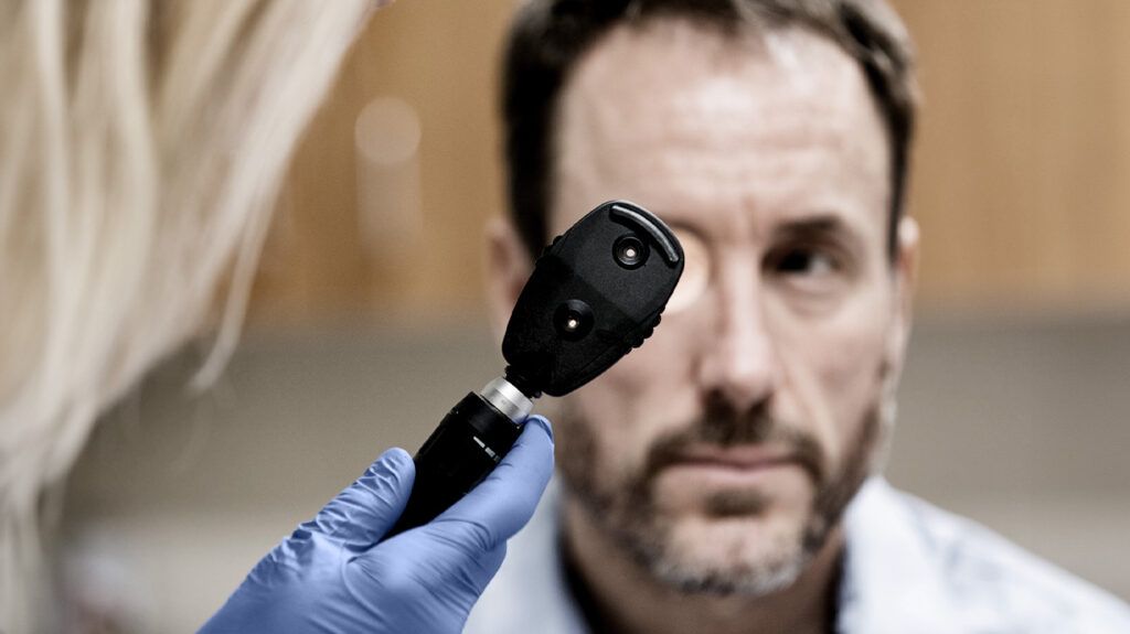
To diagnose a torn retina, an optometrist or ophthalmologist may administer eye drops to dilate the pupil. This allows them to examine the eye through a special lens called an ophthalmoscope. The doctor may also apply slight pressure to the eye.
In some cases, blood from a hemorrhage in the eye may obscure the view of the retina. If this occurs, a doctor may perform an eye ultrasound to diagnose a retinal tear.
Because the condition can progress to a retinal detachment, a person should seek urgent treatment for a retinal tear.
Retinal detachment occurs when the retina pulls away from its position in the back of the eye. This
Treatment for a retinal tear aims to prevent the condition from progressing to retinal detachment. The type of treatment a doctor uses can depend on the location of the tear.
Treatment options
Cryopexy
Cryopexy is a type of freezing treatment. It involves using a small freezing probe to form a scar around the retinal tear, helping keep it in position.
To perform cryopexy, a doctor will first numb the eye with medication. They will then touch the area of the eye nearest to the retinal tear with the freezing probe. During the procedure, a person may experience some pressure and a cold sensation.
Photocoagulation
Photocoagulation is a type of laser treatment. It involves a doctor using a laser to form small burns around the retinal tear. This creates scars to hold the retina in position.
To perform photocoagulation, the doctor will numb the eye and then shine the laser through the pupil toward the retinal tear. A person may see flashing light during the procedure.
Read about the difference between retinal tear and detachment.
Because it can occur as a natural part of the aging process, a person may not be able to prevent a retinal tear. However, a person may help reduce their risk of retinal tears and detachment by doing the
- attending regular eye exams
- using safety goggles or protective eyewear during sports or any activities that can affect the eyes
- contacting a doctor after experiencing symptoms of a retinal tear to help prevent retinal detachment
- contacting a doctor following any eye injury
A person should contact a doctor if they experience any symptoms of a retinal tear. Seeking care can help prevent the condition from progressing to retinal detachment.
Treatments for a retinal tear can also carry risks. A person who has received treatment such as cryopexy or photocoagulation should contact a doctor for any of the following:
- bleeding in the eye
- infection in the eye, which may cause swelling, discharge, and fever
- a feeling of pressure in the eye
- pain in the eye
- vision loss
With prompt diagnosis and early treatment, the outlook for a retinal tear is generally good. Because a person may develop other retinal tears in the future, continuing to attend eye appointments can help them receive appropriate monitoring and treatment.
If a retinal tear progresses to a retinal detachment, the outlook can depend on the time of diagnosis. With a timely diagnosis, treatment is successful in
Eye health resources
Visit our dedicated hub for more research-backed information and in-depth resources on eye health.
A retinal tear can occur when the receding vitreous fluid in the eye adheres to the retina and pulls it enough to cause a rip. The condition can cause floaters, flashes, and other changes in a person’s vision.
A retinal tear can progress to a retinal detachment, which can lead to permanent vision loss. A person should contact a doctor if they experience symptoms. Prompt treatment can improve outlook for someone with the condition.
Treatments for a retinal tear include freezing therapy, or cryopexy, and laser treatment, or photocoagulation.
